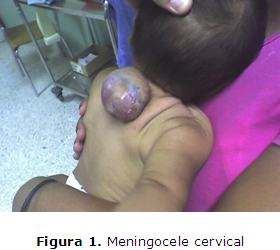
Meningocele Cervical. Presentación de un caso
Palabras clave:
Meningocele, Lactante.Resumen
El meningocele cervical es una forma rara de disrafismo espinal. La placa neural pasa por diversas transformaciones hasta convertirse en el tubo neural y cualquier alteración durante su cierre conllevará la aparición de la espina bífida. Durante el embarazo las necesidades maternas de folatos aumentan debido a la síntesis de ácidos nucleicos y proteínas durante la embriogénesis, velocidad de crecimiento y desarrollo fetal de los primeros meses de la gestación. Se presenta el caso de paciente masculino de 12 meses de edad, nacido de un parto eutócico institucional ocurrido el día 5 de diciembre del 2008, con una tumoración en la región posterior del cuello al nacer; es remitido al servicio de Neurocirugía del Hospital Mario Catarino Rivas. Se observó como dato positivo en la región cervical un aumento de volumen redondeado, adherido al plano profundo, renitente, movible y no doloroso cubierto de piel en su totalidad. Se le diagnostica un meningocele cervical y confirma mediante resonancia magnética nuclear cervicodorsal. Se realizó la resección del mismo en el mes de febrero del año 2010 y ha sido evaluado periódicamente en las consultas de neurocirugía y en su área de salud con evolución satisfactoria.
Descargas
Citas
1. Kerckoff-Villanueva HH, Bautista-Melgoza A, Rodríguez-Márquez DM. Meningocele cervical con conexión filiforme. Reporte de un caso. Ginecol Obstet Mex[internet] 2011[citado enero 2012];79(8):497-500. Disponible en: http://www.nietoeditores.com.mx/download/gineco/2011/AGOSTO2011/FEMEGO8.11MENINGOCELE.pdf
2. Tungaria A, Kumar Srivastav A, Mahapatra AK, Kumar R. Multiple neural tube defects in the same patient with no neurological déficit. J Pediatr Neurosci[internet]. 2010 Jan-Jun[cited january 2012]; 5(1): 52-54. Available from: http://www.ncbi.nlm.nih.gov/pmc/articles/PMC2964786/
3. Rivero-Celada D, Carcavilla-Loncán LI, Marín-Cárdenas MA, Cantero-Antón JM, Alfaro-Torres J, Duato-Jané F, et al. Degeneración tumoral en meningocele no intervenido. Descripción de dos casos. Neurocirugía[internet]. 2006[citado enero 2012]; 17(6): 532-537. Disponible en: http://scielo.isciii.es/pdf/neuro/v17n6/4.pdf
4. Senoglu M, Yilmaz Z. Cervical meningocele with tethered cord in a seven -years old child: Case Report. The Internet Journal of Pediatrics and Neonatology[internet]. 2009[cited january 2012];10(1): Available from: http://www.ispub.com/journal/the-internet-journal-of-pediatrics-and-neonatology/volume-10-number-1/cervical-meningocele-with-tethered-
cord-in-a-seven-years-old-child-case-report.html
5. Tarqui-Mamani C, Sanabria H, Lam N, Arias J. Incidencia de los defectos del tubo neural en el Instituto Nacional Materno Perinatal de Lima. Rev Chil Salud Pública[internet] 2009[citado enero 2012];13(2): 82-89. Disponible en: http://www.revistasaludpublica.uchile.cl/index.php/RCSP/article/viewFile/614/518
6. Eller TW, Bernstein LP, Rosenberg RS, McLone DG. Tethered cervical spinal cord. Case report. J Neurosurg. 1987;67:600-2.
7. Iizuka T. Fatty Filum Terminale on MRI. The Internet Journal of Spine Surgery[ìnternet]. 2007[cited january 2012];3(1): Available from: http://www.ispub.com/journal/the-internet-journal-of-spine-surgery/volume-3-number-1/fatty-filum-terminale-on-mri.html
8. Sáez Martín A, Moreno C, Platas M, Lambre J, Bernachea J, Landaburu P. Dilatación del ventrículo terminal: Presentación de un caso. Revisión de la literatura. Rev. argent. neurocir. [revista en la Internet]. 2007 Sep [citado 2012 Feb 22]; 21(3): Disponible en: http://www.scielo.org.ar/scielo.php?script=sci_arttext&pid=S1850-15322007000300014&lng=es.
9. Shane Tubos R, Oakes WJ. A simple method to deter retethering in patients with spinal dysraphism[technical note]. Childs Nerv Syst[internet].2006[cited january 2012];22: 715-716. Available from: http://www.springerlink.com/content/946528722782474m/fulltext.pdf
10. Martínez-Lage JF, Ruiz-Espejo Vilar A, Almagro MJ, Sánchez del Rincón I, Ros de San Pedro J, Felipe-Murcia M. et al. Reanclaje medular en pacientes con mielomeningocele y lipomeningocele: la segunda operación. Neurocirugía [revista en la Internet]. 2007 Ago [citado 2012 Feb 22]; 18(4): 312-319. Disponible en: http://scielo.isciii.es/scielo.php?script=sci_arttext&pid=S1130-14732007000400004&lng=es.
11. Brezner A, Kay B. Spinal cord ultrasonography in children with myelomeningocele. Dev Med Child Neurol. 1999;41:450_455 http://onlinelibrary.wiley.com/doi/10.1111/j.1469-8749.1999.tb00637.x/pdf
12. Fong D. Spinal Dysraphism. The Hong Kong Medical Diary[internet]. december 2006[cited january 2012]; 11(12):Available from: http://www.fmshk.org/database/articles/mb02drdawsonfong.pdf
13. Bulent Düz, Selcuk Gocmen, Halil Ibrahim Secer, Seref Basal, Engin Gönül. Tethered Cord Syndrome in Adulthood J Spinal Cord Medìnternet]. 2008[cited january 2012]; 31(3): 272-278. Available from: http://www.ncbi.nlm.nih.gov/pmc/articles/PMC2565560/
14. Myeong Jin Kim, Soo Han Yoon, Ki Hong Cho, Geun Soo Won. Tethered Spinal Cord with Double Spinal Lipomas. Korean Med Sciìnternet]. 2006 December[cited january 2012]; 21(6): 1133-1135. Available from: http://www.ncbi.nlm.nih.gov/pmc/articles/PMC2721945/
15. Aguiar CA, Mendoza-Lattes S, Cobb P, Menezes A, Weinstein SL. Unusual Association of Congenital Kyphosis and Conus Lipoma Presenting as a Double Spinal Cord Tether Iowa Orthop J[internet]. 2007[cited january 2012]; 27: 85-89. Available from: http://ukpmc.ac.uk/articles/PMC2150652/pdf/iowa0027-0085.pdf
16. Reigel DH, Tchernoukha K, Bazmi B, et al. Change in spinal curvature following release of tethered spinal cord associated with spina bifida. Pediatr Neurosurg. 1994; 20:30-42.
17. Ramesh VV Ch. Phani MK. Cervical myelocystocele: Case report and review of literature. J Pediatr Neurosci[internet]. 2011 Jan-Jun[cited january 2012]; 6(1): 55-57. Available from: http://www.ncbi.nlm.nih.gov/pmc/articles/PMC3173918/
18. Herman JM, McLone DG, Storrs BB, et al. Analysis of 153 patients with myelomeningocele or spinal lipoma reoperated upon for a tethered cord. Presentation, management, and outcome. Pediatr Neurosurg.1993; 19(5):243-49.

Publicado
Cómo citar
Número
Sección
Licencia
Aquellos autores/as que tengan publicaciones con esta revista, aceptan los términos siguientes:- Los autores/as conservarán sus derechos de autor y garantizarán a la revista el derecho de primera publicación de su obra, el cuál estará simultáneamente sujeto a la Licencia de reconocimiento de Creative Commons que permite a terceros compartir la obra siempre que se indique su autor y su primera publicación esta revista.
- Los autores/as podrán adoptar otros acuerdos de licencia no exclusiva de distribución de la versión de la obra publicada (p. ej.: depositarla en un archivo telemático institucional o publicarla en un volumen monográfico) siempre que se indique la publicación inicial en esta revista.
- Se permite y recomienda a los autores/as difundir su obra a través de Internet (p. ej.: en archivos telemáticos institucionales o en su página web) antes y durante el proceso de envío, lo cual puede producir intercambios interesantes y aumentar las citas de la obra publicada. (Véase El efecto del acceso abierto).


