Transformation of hyperplastic polyps to ulcerated mucinous adenocarcinoma of the colon
Keywords:
COLON, MUCINOUS ADENOCARCINOMA, RADIOGRAPHY.Abstract
Introduction: colorectal cancer includes any type of colon cancer, rectum and appendix and many cases have their origin in an adenomatous polyp.
Objective: to describe the evolution of hyperplastic polyps to mucinous colon cancer through the analysis of a case where the imaging study contributed to its diagnosis and an accurate surgery.
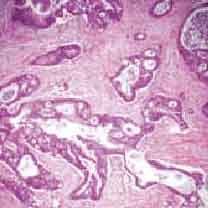
Case report: 72 year-old female patient, who has been treated since 2012 because of hyperplastic colon polyps and chronic colitis. In 2014 she presented abdominal pain in the right flank, vomiting and diarrhea. Colonoscopy confirmed the existence of hyperplastic polyps in the cecum and rectum. The abdominal ultrasound scan showed a complex and solid mass in the hypochondrium and right flank, very close to the peritoneum, impressing a tumor lesion of the digestive tract. Intestinal transit radiography showed a filling defect in the cecum, infiltrating the ileocecal valve. In simple and proven multislice tomography performed by oral and intravenous route a tumor lesion occluding the light of cecum was observed, with hypercaptation of contrast in both phases. The colonoscopy confirmed an injury to the ileocecal valve and another at ascending colon level, both of dubious appearance and rectosigmoid polyps. Colonoscopy biopsy confirmed an ulcerated and infiltrating mucinous adenocarcinoma.
Conclusions: it has been proved that one of the origins of mucinous adenocarcinomas of colon originates from benign polyps and imaging studies are a useful tool in the staging of the tumor and the treatment to be followed.
COLON; MUCINOUS ADENOCARCINOMA; RADIOGRAPHY.
Downloads
References
1. Siegel R, Ma J, Zou Z, Jemal A. Cancer statistics, 2014. CA Cancer J Clin [Internet] 2014; 64(1): 9-29. Disponible en: http://onlinelibrary.wiley.com/doi/10.3322/caac.21208/full
2. SINAIS/SINAVE/DGE/SALUD. Perfil epidemiológico de los tumores malignos en México 2011. México [Internet]: Secretaría de Salud; 2011. Disponible en: http://docplayer.es/11449544-Perfil-epidemiologico-de-los-tumores-malignos-en-mexico.html
3. Gao P, Song YX, Xu YY, Sun Z, Sun JX, Xu HM, et al. Does the prognosis of colorectal mucinous carcinoma depend upon the primary tumour site? Results from two independent databases. Histopathology [Internet] 2013; 63(5):603-15. Disponible en: https://www.researchgate.net/publication/256328074_Does_the_prognosis_of_colorectal_mucinous_carcinoma_depend_upon_the_primary_tumour_site_Results_from_two_independent_databases
4. Mantilla Morales A, Mendoza Morales RC, Alvarado Cabreroc I. Evaluación de piezas quirúrgicas con carcinoma de colon. Gaceta Mexicana de Oncología. [Internet] 2014;13(4):229-35. Disponible en: http://www.google.com.cu/url?sa=t&rct=j&q=&esrc=s&source=web&cd=1&cad=rja&uact=8&ved=0ahUKEwiByuaEsO3SAhUr0YMKHeaqD5kQFggYMAA&url=http%3A%2F%2Fwww.elsevier.es%2Fes-revista-gaceta-mexicana-oncologia-305-pdf-X166592011457905X-S300&usg=AFQjCNEeARbDXTcr-8R8UtvEjNN4V2F-gg&bvm=bv.150475504,d.amc
5. Dirección de registros médicos y estadísticas de salud. Estadísticas de salud en Cuba-Anuario Estadístico 2014 [Internet]. La Habana Cuba, 2015 [Consultado 06 ene 2016]. Disponible en: http://www.google.com.cu/url?sa=t&rct=j&q=&esrc=s&source=web&cd=1&cad=rja&uact=8&ved=0ahUKEwjRmeWUsu3SAhWG34MKHUcjCxcQFggYMAA&url=http%3A%2F%2Ffiles.sld.cu%2Fbvscuba%2Ffiles%2F2015%2F04%2Fanuario-estadistico-de-salud-2014.pdf&usg=AFQjCNH1MiNBJ-L9aet6CAKuN7xk8YHTQA&bvm=bv.150475504,d.amc
6. Bosch SL, Teerenstra S, de Wilt JH, Cunningham C, Nagtegaal ID. Predicting lymph node metastasis in pT1 colorectal cancer: a systematic review of risk factors providing rationale for therapy decisions. Endoscopy 2013 [Internet]; 45(10): 827-834. Disponible en: https://www.ncbi.nlm.nih.gov/pubmed/23884793
7. Bannura Cumsille G. ¿Se puede mejorar la oportunidad del diagnóstico del cáncer colorrectal en Chile? Rev. Chil. Cir. [Internet] 2006; 58(1):59-61. Disponible en: http://www.scielo.cl/scielo.php?script=sci_arttext&pid=S0718-40262006000100014
8. Minoo P, Zlobec I, Peterson M, Terracciano l, Lugli A. Characterization of rectal, proximal and distal colon cancers based on clinicopathological, molecular and protein profiles. Int J of Onc. [Internet] 2010; 37(3): 707-18. Disponible en: https://www.ncbi.nlm.nih.gov/pubmed/20664940
9. Verhulst J, Ferdinande L, Demetter P, Ceelen W. Mucinous subtype as prognostic factor in colorectal cancer: a systematic review and meta-analysis. J Clin Pathol. [Internet] 2012; 65(5):381-8. Disponible en: http://jcp.bmj.com/content/65/5/381.long
10. Zerhouni EA, Rutter C, Hamilton SR, Balfe DM, Megibow AJ, Francis IR, et al. CT and MR imaging in the staging of colorectal carcinoma: report of the Radiology Diagnostic Oncology Group II. Radiology [Internet] 1996, 200(2): 443-51. Disponible en: http://pubs.rsna.org/doi/pdf/10.1148/radiology.200.2.8685340
11. García I. Estudio de la supervivencia en el cáncer colorectal en relación con el grado arquitectural sumatorio y topográfico. [Tesis de Doctorado] [Internet]. Universidad de Málaga, España. 2013. Disponible en: http://www.google.com.cu/url?sa=t&rct=j&q=&esrc=s&source=web&cd=1&cad=rja&uact=8&ved=0ahUKEwjd5qaYru_SAhUL0oMKHS2ODWAQFggbMAA&url=http%3A%2F%2Fwww.riuma.uma.es%2Fxmlui%2Fbitstream%2Fhandle%2F10630%2F6460%2FTDR_GARCIA_MUIOZ.pdf%3Fsequence%3D1&usg=AFQjCNGXnPfcmeKF-cRZLnXEWYnJHkN2Hg&bvm=bv.150475504,d.eWE
12. Artega R, Boscán A, Quero R, Rojas N, Sposito F. Cáncer de colon y recto en pediatría. Presentación de casos. Rev Venez Oncol [Internet]. 2013; 25(2):104-8. Disponible en: http://www.google.com.cu/url?sa=t&rct=j&q=&esrc=s&source=web&cd=1&cad=rja&uact=8&ved=0ahUKEwih4ImdsO_SAhXoxYMKHTXGB1cQFggYMAA&url=http%3A%2F%2Fwww.oncologia.org.ve%2Fsite%2Fupload%2Frevista%2Fpdf%2F07.__arteaga_r_%2528104-108%2529.pdf&usg=AFQjCNFl6kFFdlYzmFb8pX8-pwHCsMgStQ&bvm=bv.150475504,d.eWE

Published
How to Cite
Issue
Section
License
Authors who have publications with this journal agree to the following terms: Authors will retain their copyrights and grant the journal the right of first publication of their work, which will be publication of their work, which will be simultaneously subject to the Creative Commons Attribution License (CC-BY-NC 4.0) that allows third parties to share the work as long as its author and first publication in this journal are indicated.
Authors may adopt other non-exclusive license agreements for distribution of the published version of the work (e.g.: deposit it in an institutional telematic archive or publish it in a volume). Likewise, and according to the recommendations of the Medical Sciences Editorial (ECIMED), authors must declare in each article their contribution according to the CRediT taxonomy (contributor roles). This taxonomy includes 14 roles, which can be used to represent the tasks typically performed by contributors in scientific academic production. It should be consulted in monograph) whenever initial publication in this journal is indicated. Authors are allowed and encouraged to disseminate their work through the Internet (e.g., in institutional telematic archives or on their web page) before and during the submission process, which may produce interesting exchanges and increase citations of the published work. (See The effect of open access). https://casrai.org/credit/


