

Phacoemulsification in patients with primary angular closure
Keywords:
PHACOEMULSIFICATION; CATARACT EXTRACTION; INTRAOCULAR PRESSURE; ANTERIOR CHAMBER.Abstract
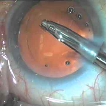
Introduction: there is currently a tendency toward phacoemulsification in the primary angular closure due to the crucial role of the lens in the pathophysiology of the disease.
Objective: to describe the results of phacoemulsification in patients with primary angular closure in Pinar del Río.
Method: a prospective longitudinal descriptive study was carried out in Pinar del Río, between January 2013 and December 2015, which included 43 patients with primary angular closure not controlled by laser peripheral iridotomy or medical treatment, who underwent phacoemulsification. Statistical analysis was performed using the SPSS program.
Results: mean age was 54.67 ± 4.22 years and 83.7% were women. The mean amplitude value of the anterior chamber was 2.56 0.11 mm increasing to 3.24 0.08 mm (p = 0.006). 93.02% of the eyes showed a narrow angle, and after treatment 83.72% presented a grade 3-4 (p <0.001). The best corrected average visual acuity varied from 0.74 to 0.98 (p = 0.042) and the refractive sphere from 3.25 to -0.43 diopters (p = 0.001). The intraocular pressure was modified from 26.7 ± 3.3 mmHg in the preoperative period to 15.1 ± 1.8 mmHg 6 months after surgery (p <0.001). The transoperative and postoperative complications were present in 2.3% and 4.7%, respectively.
Conclusion: the results of phacoemulsification in patients with primary angular closure were satisfactory since an improvement in anatomic and refractory parameters was observed, with intraocular pressure control and a minimum of complications.
Downloads
References
1. Fernández Argones L, Piloto Díaz I, Díaz Águila Y, Obret Mendive I, Ferrer Guerra MT, Álvarez Cisneros G. Consideraciones terapéuticas en el cierre angular primario. Rev Cubana Oftalmol [Internet]. 2012 [consultado 15 Nov 2016]; 25(1). Disponible en: http://scielo.sld.cu/scielo.php?script=sci_arttext&pid=S0864-21762012000300009&lng=es
2. Pérez González H, García Concha Y, Moreno Domínguez JC, Gómez Martínez N. Opciones quirúrgicas en el cierre angular primario por iris en meseta. Rev Cubana Oftalmol [Internet]. 2016 [consultado 15 Nov 2016]; 29(1). Disponible en: http://www.revoftalmologia.sld.cu/index.php/oftalmologia/article/view/298
3. Foster PJ, Buhrmann R, Quigley HA, et al. The definition and classification of glaucoma in prevalence surveys. Brit J Ophthalmol [Internet]. 2002 [consultado 21 Nov 2016]; 86(2):238-42. Disponible en: http://bjo.bmj.com/content/86/2/238.full
4. Pérez-González H, Garcia-Concha Y, Gómez–Martínez N. Cierre angular primario: opciones quirúrgicas. Rev Mex Oftalmol. 2014 [consultado 21 Nov 2016]; 88(4):182-85. Disponible en: http://www.sciencedirect.com/science/article/pii/S0187451914000547
5. Piloto Díaz I, Río Torres I, Fernández Argones L, Barroso Lorenzo R. Facoemulsificación en el Cierre Angular Primario Agudo. En: Centurion V, Nicoli C, Chávez E (ed). Cristalino de las Américas: La Cirugía del Cristalino Hoy. 2da ed. Panamá: Jaypee-Highlights; 2015. p. 959-65.
6. Muniesa Royo MJ. Facoemulsificación en el glaucoma crónico de ángulo cerrado. Annals d’Oftalmologia [Internet]. 2015 [consultado 3 Dic 2016]; 23(4):320-27. Disponible en: http://www.annalsoftalmologia.com/articulos/a18657/08_1_dra-muniesa.pdf
7. Biblioteca Nacional de Medicina de los EE.UU. Glaucoma. Medline Plus [Internet] 2015. Disponible en: https://medlineplus.gov/spanish/ency/article/001620.htm
8. Man X, Chan NC, Baig N, Kwong Y, Leung D, Li F. Anatomical effects of clear lens extraction by phacoemulsification versus trabeculectomy on anterior chamber drainage angle in primary angle-closure glaucoma (PACG) patients. Graefes Arch Clin Exp Ophthalmol [Internet]. 2015 [consultado 5 Dic 2016]; 253(5):773–78. Disponible en: http://link.springer.com/article/10.1007/s00417-015-2936-z
9. Nongpiur ME, Haaland BA, Perera SA, Fried DS, Sakata LM et al. Development of a score and probability estimate for detecting angle closure based on anterior segment optical coherence tomography. Am J Ophthalmol [Internet]. 2014 [consultado 5 Dic 2016]; 157(1):32-8. Disponible en: http://www.sciencedirect.com/science/article/pii/S0002939413006193
10. Mösler MP, Werner JU, Lang GK. Chamber angle assessment in clinical practice, a comparison between optical coherence tomography and gonioscopy. Klinische Monatsblatter Fur Augenheilkunde. 2015 [consultado 22 Dic 2016] 232(7):874-880. Disponible en: http://europepmc.org/abstract/med/25462938
11. Romera Romero P, Carbonell Puig M, Moll Udina A, Romanic Bubalo N, Castellvi Manent J. Utilidad de la tomografía óptica del segment anterior (OCT-SA) en el glaucoma. Annals d’Oftalmologia [Internet]. 2015 [consultado 27 Dic 2016]; 23(4):27-33. Disponible en: http://www.annalsoftalmologia.com/articulos/a18618/02_3_dr-romera.pdf
12. Castany Aregall M, Rigo Quera J, Carceller Guillamet, Pujadas García, Dou Sáenz de Vizmanos A. Goniosinequiólisis. Annals d’Oftalmologia [Internet]. 2015 [consultado 27 Dic 2016]; 23(4):341-49. Disponible en: http://www.annalsoftalmologia.com/articulos/a18659/08_3_dra-castany.pdf
13. Dada T, Rathi A, Angmo D, Agarwal T, Vaqnathi M, Khokhar S. Clinical outcomes of clear lens extraction in eyes with primary angle closure. Journ Cataract & Refract Surg [Internet]. 2015 [consultado 27 Dic 2016]; 41(7):1470-77. Disponible en: http://dx.doi.org/10.1016/j.jcrs.2014.10.029
14. Jarrín E, Cabarga-Nozal C, Almendral A, Muñoz-Negrete FJ. Iridotomía periférica láser frente a facoemulsificación en el cierre angular primario: estudio comparativo prospectivo. Arch Soc Esp Oftalmol [Internet]. 2014 [consultado 27 Dic 2016]; 89(9):352–60. Disponible en: http://www.sciencedirect.com/science/article/pii/S0365669114001476
Published
How to Cite
Issue
Section
License
Authors who have publications with this journal agree to the following terms: Authors will retain their copyrights and grant the journal the right of first publication of their work, which will be publication of their work, which will be simultaneously subject to the Creative Commons Attribution License (CC-BY-NC 4.0) that allows third parties to share the work as long as its author and first publication in this journal are indicated.
Authors may adopt other non-exclusive license agreements for distribution of the published version of the work (e.g.: deposit it in an institutional telematic archive or publish it in a volume). Likewise, and according to the recommendations of the Medical Sciences Editorial (ECIMED), authors must declare in each article their contribution according to the CRediT taxonomy (contributor roles). This taxonomy includes 14 roles, which can be used to represent the tasks typically performed by contributors in scientific academic production. It should be consulted in monograph) whenever initial publication in this journal is indicated. Authors are allowed and encouraged to disseminate their work through the Internet (e.g., in institutional telematic archives or on their web page) before and during the submission process, which may produce interesting exchanges and increase citations of the published work. (See The effect of open access). https://casrai.org/credit/
