

Meningioma of the optic nerve sheaths
Keywords:
MENINGIOMA; OPTIC NERVE; OPTIC NERVE NEOPLASMS.Abstract
Introduction: meningiomas of the optic nerve sheath are meningothelial tumors originating from arachnoid cells that produce decrease of vision and proptosis with variable ocular involvement.
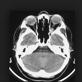
Case report: a 65-year-old female patient with a history of hypertension who was admitted due to proptosis, decrease of vision and conjunctival chemosis of the right eye; fundus of the eye examination revealed the presence of a small optic disc of defined edges of normal coloration, without compromise of the thickness of the layer of nervous fibers of the retina with moderate vascular dilatation to venous predominance without evidence of macular alterations, hemorrhages or exudates. The campimetric studies showed the presence of altitudinal scotoma with lower temporal quadrant capture, and the study of the visual evoked potentials showed fall of its amplitude. Through the study of nuclear magnetic resonance (NMR, the existence of a homogeneous isointense image of well-defined edges was observed, which did not compromise the axonal structure of the optic nerve and which respects the intracanalicular and intracranial portion that confirmed the diagnosis of meningioma of the optic nerve sheaths.
Discussion: the diagnosis of meningioma of the optic nerve sheaths on the right eye was confirmed in a patient with unilateral proptosis and conjunctival chemosis, which constitutes an unusual presentation with ophthalmological manifestations.
Downloads
References
1. Leiria de Moura da Silva L, Barbosa-Coutinho LM. Processos expansivos orbitoesfenoidais: estudo anatomopatológico de 82 casos. Arq. Bras. Oftalmol. [Internet]. 2009 Feb [cited 2017 Dec 08] ; 72( 1 ): 84-90. Disponible en: http://www.scielo.br/scielo.php?script=sci_arttext&pid=S0004-27492009000100017&lng=en.
2. Srikant S. Histopathological analysis of meningioma and its variants: A study of fifty cases. Indian Journal of Cancer. [Internet]. 2017[cited 2017 Dec 08];54(1):313-5 Disponible en: http://www.indianjcancer.com/temp/IndianJournalofCancer541313-2621357_071653.pdf
3. Peyre M, Feuvret L, Sanson M, Navarro S, Boch A L, Loiseau H, et al. Diffuse midline skull base meningiomas: identification of a rare and aggressive subgroup of meningiomas. Journal of Neuro Oncology. 2017;133(3):633–9. Disponible en: https://www.ncbi.nlm.nih.gov/pubmed/28536991.
4. Muci Mendoza R. Veinticinco años en la Unidad de Neuro-oftalmología del Hospital Vargas de Caracas.(1980-2005). Gav Méd Caracas. 2007; 115(4) 313’324. Disponible en: https://www.bitacoramedica.com/wp-content/uploads/2013/02/Seccion-10-MUCI-MENDOZA-25-an%C2%A6%C3%A2os-de-la-Unidad-de-Neurooftalmologia-del-Hospital-Vargas.pdf
5. Nguema Afumu C. Rodríguez Ramos JF, Rodríguez Villalonga OL, Arenas Rodríguez I, Boffill Corrales AM. Los meningiomas intracraneales recidivantes postquirúrgicos. Rev Ciencias Médicas [Internet]. 2014 Abr [citado 2017 Dic 08] ; 18( 2 ): 231-243. Disponible en: http://scielo.sld.cu/scielo.php?script=sci_arttext&pid=S1561-31942014000200007&lng=es
6. Meeker A R, Ko M W, Carruth B P, Strumpf K B, Bersani T A. Diagnosis of optic nerve sheath meningioma during optic nerve sheath decompression. The International Journal on Orbital Disorders, Oculoplastic and Lacrimal Surgery. 2017;36(1):35-8. Disponible en: https://www.ncbi.nlm.nih.gov/pubmed/28156180.
7. Hokazono K, Moura Castelo F, Monteiro Ribeiro M L. Meningioma do nervo óptico simulando progressão de dano axonal glaucomatoso: relato de caso. Arq. Bras. Oftalmol. [Internet]. 2008 Oct [cited 2017 Dec 08] ; 71( 5 ): 725-728. Disponible en: http://www.scielo.br/scielo.php?script=sci_arttext&pid=S0004-27492008000500023&lng=en. http://dx.doi.org/10.1590/S0004-27492008000500023.
8. Hoch M J, Bruno M T, Shepherd T M. Advanced MRI of the Optic Nerve. Journal of Neuro-Ophthalmology 2017;37(2):187–96
9. Caeiro M, Conde C, López ML, Pérez L, Vázquez de la Torre ML, Canteli M et al. Meningioma de la vaina del nervio óptico (MVNO). ¿La radioterapia es el actual patrón de cuidados?: a propósito de un caso y revisión de la literatura. Oncología (Barc.) [Internet]. 2006 Ene [citado 2017 Dic 08] ; 29( 1 ): 38-46. Disponible en: http://scielo.isciii.es/scielo.php?script=sci_arttext&pid=S0378-48352006000100005&lng=es.
10. Jayanetti V, Klistorner A I, Graham S L, Dexter M, Flaherty M P, Jones K, et al. Monitoring of optic nerve function in Neurofibromatosis 2 children with optic nerve sheath meningiomas using multifocal visual evoked potentials. Journal of Clinical Neuroscience 2018; 50: 262-267. Disponible en: https://www.sciencedirect.com/science/article/pii/S096758681731233X?via%3Dihub.
Published
How to Cite
Issue
Section
License
Authors who have publications with this journal agree to the following terms: Authors will retain their copyrights and grant the journal the right of first publication of their work, which will be publication of their work, which will be simultaneously subject to the Creative Commons Attribution License (CC-BY-NC 4.0) that allows third parties to share the work as long as its author and first publication in this journal are indicated.
Authors may adopt other non-exclusive license agreements for distribution of the published version of the work (e.g.: deposit it in an institutional telematic archive or publish it in a volume). Likewise, and according to the recommendations of the Medical Sciences Editorial (ECIMED), authors must declare in each article their contribution according to the CRediT taxonomy (contributor roles). This taxonomy includes 14 roles, which can be used to represent the tasks typically performed by contributors in scientific academic production. It should be consulted in monograph) whenever initial publication in this journal is indicated. Authors are allowed and encouraged to disseminate their work through the Internet (e.g., in institutional telematic archives or on their web page) before and during the submission process, which may produce interesting exchanges and increase citations of the published work. (See The effect of open access). https://casrai.org/credit/
