

Ewing's sarcoma of retroperitoneal presentation
Keywords:
NEUROECTODERMAL TUMORS; PRIMITIVE; PERIPHERAL/diagnostic; SARCOMA; EWING/diagnostic; IMMUNOHISTOCHEMISTRY; BIOPSY; FINE-NEEDLE; MICROSCOPY.Abstract
Introduction: within the tumors of small, round, blue cells are a heterogeneous range of neoplasm in which immunohistochemical or other special techniques are imperative to be able to differentiate, or in many cases, only by exclusion approaching the possible histogenesis of the tumor in any case, these tumors are all highly undifferentiated, aggressive and almost always of a gloomy or very poor prognosis.
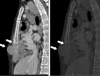
Case Report: a 44-year-old male patient, with pathological history of acute lymphoblastic leukemia, with a complete remission of 10 year-evolution, who was assessed by presenting pain in the left lumbar region. A case study was performed and ultrasound revealed the presence of a retroperitoneal tumor. Surgery was decided on by exploratory laparotomy, and surgical excision of the tumor was performed. The patient recovery was satisfactory from the surgical procedure, however, the tumor recurred in a period of 1 month and the treatment with radiotherapy and cytostatics started with an unfavorable prognosis.
Conclusions: the definitive diagnosis was performed by immunohistochemical study, resulting in a peripheral neuroectodermal tumor. The definitive diagnosis is made by the immunohistochemical study, the result being a peripheral neuroectodermal tumor or Ewing's sarcoma. Given that it is a rare diagnosis in adults after the second decade of life and often confused with other tumors, such as melanomas or carcinomas, the case is reported.
Downloads
References
1. Briseño AA, Quesada DR, Corona E, Castañeda A, Duarte A, Duarte, Dassaejv M. Tumor intraabdominal desmoplásico de células pequeñas y redondas. Cirugía y cirujanos [en línea]. 2015 [Citado 21 febrero 2017]; 83(3): [5p.]. Disponible en: https://www.sciencedirect.com/science/article/pii/S0009741115000675
2. Mosquera Betancourt G, Hernández González EH, Hernández Cabezas I, Quintero Martínez O. Sarcoma de Ewing extraesquelético del raquis dorsal: presentación de dos casos. Rev Cubana Neurol Neurocir. [en línea]. 2014 [Citado 21 febrero 2017]; 4(2): [7p.]. Disponible en: http://www.medigraphic.com/pdfs/revcubneuro/cnn-2014/cnn142n.pdf
3. Ríos L, Vásquez L, Silva JM, Sialer L, Maza I, Oscanoa M, et al. Prognostic factors and survival in patients under 18 years of age with Ewing sarcoma family tumors: a 10-year experience. Horiz. Med. [en línea]. 2017 Oct [Citado 2018 Feb 23]; 17(4): [8p.]. Disponible en: http://www.scielo.org.pe/scielo.php?script=sci_arttext&pid=S1727-558X2017000400002&lng=es
4. Álvarez San Nicolás J. Cirugía de salvamento de extremidad en sarcoma de Ewing. 2015. [Tesis Doctoral]. Barcelona: Universidad Autónoma. Facultad de Medicina; 2015 [citado 18 Feb 2017]. 381 p. Disponible en: https://ddd.uab.cat/pub/tesis/2015/hdl_10803_310613/jasn1de1.pdf
5. Gallegos Castorena S, Cárdenas Cardos R. Sarcoma de Ewing y tumor neuroectodérmico primitivo: Protocolos técnicos cáncer en niños. México, D.F.: Editores de textos mexicanos S.A. de C.V; 2010. 219-33p.
6. Villalta Fallas JC. Sarcoma de Ewing. Revista Médica de Costa Rica y Centroamérica [en línea]. 2016 [citado 2018 Feb 23]; 72(617): [9p.]. Disponible en: http://www.medigraphic.com/pdfs/revmedcoscen/rmc-2015/rmc154c.pdf
7. Riveros Ramos LC, Velasco Hidalgo L, Shalkow Klincovstein J, Rojas Maruri CM, Cárdenas Cardos R, Rivera Luna R. Tumor neuroectodérmico primitivo primario de páncreas en un paciente pediátrico. Reporte de caso y revisión de la literatura. Acta Pediatr Mex. [en línea]. 2016 [citado 2018 Feb 23]; 37(1): [5p.]. Disponible en: http://www.scielo.org.mx/pdf/apm/v37n1/2395-8235-apm-37-01-00026.pdf
8. Soto C, Gómez LC, Criollo F, Romo R, Messa O, Arbeláez P. Sarcoma de Ewing de la falange proximal del meñique. Reporte de caso. Rev. Colomb. Cancerol. [Internet]. 2014 Sep [cited 2018 Feb 23]; 18(3): [5p.] Available from: http://www.scielo.org.co/scielo.php?script=sci_arttext&pid=S0123-90152014000300006&lng=en
9. Fernández A, Perfetti W, Tellez R, Scarton J, Verdecchia D, Sarmiento P, et al. Condrosarcoma mesenquimático extraesquelético de región cervical. Rev Venez de Oncol [Internet] 2015 [citado 2018 Feb 23]; 27(1): [5p.]. Disponible en: http://www.redalyc.org/articulo.oa?id=375637056008
10. Benjamín RS. Bone Sarcomas. In: Skeel RT, ed. Handbook of Cancer Chemoterapy. 7th ed. New York: Lippinkott Williams & Wilkins; 2007. p. 434-40.
Published
How to Cite
Issue
Section
License
Authors who have publications with this journal agree to the following terms: Authors will retain their copyrights and grant the journal the right of first publication of their work, which will be publication of their work, which will be simultaneously subject to the Creative Commons Attribution License (CC-BY-NC 4.0) that allows third parties to share the work as long as its author and first publication in this journal are indicated.
Authors may adopt other non-exclusive license agreements for distribution of the published version of the work (e.g.: deposit it in an institutional telematic archive or publish it in a volume). Likewise, and according to the recommendations of the Medical Sciences Editorial (ECIMED), authors must declare in each article their contribution according to the CRediT taxonomy (contributor roles). This taxonomy includes 14 roles, which can be used to represent the tasks typically performed by contributors in scientific academic production. It should be consulted in monograph) whenever initial publication in this journal is indicated. Authors are allowed and encouraged to disseminate their work through the Internet (e.g., in institutional telematic archives or on their web page) before and during the submission process, which may produce interesting exchanges and increase citations of the published work. (See The effect of open access). https://casrai.org/credit/
